Researchers have developed a simple method to simultaneously acquire images at different depths with a standard microscope. The new technique can be applied to a variety of microscopy methods, making it useful for a variety of biological and biomedical imaging applications.
“Optical microscopy has been an indispensable tool for studying complex 3D biological systems and processes,” said Sheng Xiao, a member of the Boston University research team. “With our new multifocus technology, living cells and organisms can be observed at high speed and high contrast.”
Im opticsResearchers from the Optical Society (OSA) for Highly Effective Research, led by Jerome Mertz, describe their new simple and fast way to get information from different depths using standard microscopy. The new approach can be easily added to most existing systems and is easy to replicate so that it is accessible to other researchers.
Taking multi-focus images
Standard camera-based microscopy systems capture sharp images in a single focal plane. Although researchers have tried different strategies to simultaneously capture images with different focal lengths, these approaches typically require multiple cameras or use a special diffractive optical element to perform the image division with a single camera. Both strategies are complex and it can be difficult to make a diffractive optical element.
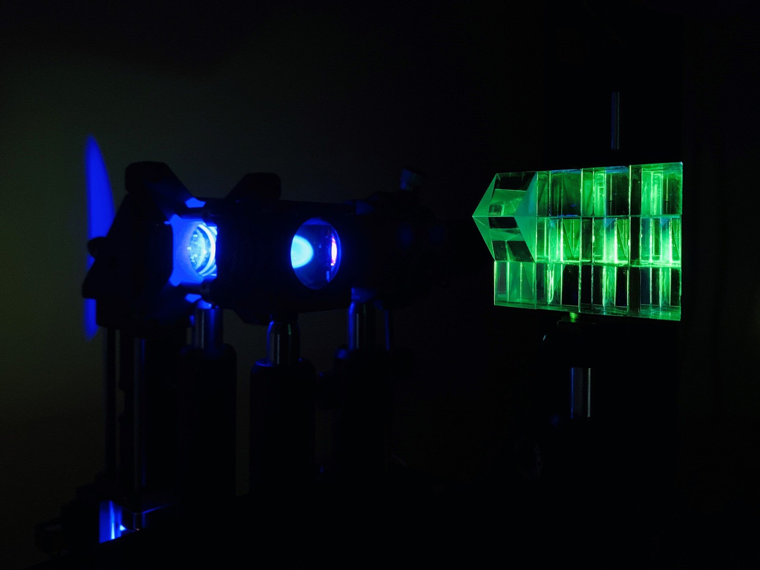
“We used a Z-splitter prism that can be assembled entirely from off-the-shelf components and easily applied to a variety of imaging modalities such as fluorescence, phase contrast, or dark field imaging,” said Xiao.
The Z-splitter prism splits detected light to produce multiple images simultaneously in a single camera frame. Each image is focused to a different depth in the sample. Using a high-speed camera with a large sensor area and a high number of pixels allowed the researchers to distribute multiple high-resolution images without overlapping on the same sensor.
The multifocal images recorded with the new technology make it possible to estimate the blurred background from the sample much more precisely than is possible with a single image. Researchers used this information to develop an improved 3-D deburring algorithm that eliminates the blurry background light that is often a problem when using wide-field microscopy.
“Our 3D deblurring extended volume algorithm suppresses blurred background from sources outside the image volume,” said Xiao. “This improves both the image contrast and the signal-to-noise ratio, which makes it particularly advantageous in fluorescence imaging applications with thick samples.”
Proven versatility
Researchers demonstrated the new technique using commonly used microscopy modalities, including fluorescence, phase contrast, and dark field imaging. They took large field-of-view 3D images that spanned hundreds of neurons or entire freely moving organisms, as well as high-speed 3D images of a rotifer cilia beating every hundredth of a second. This demonstrated how the approach offers the flexibility to prioritize a large field of view or high speed.
To demonstrate the capabilities of the 3D deblurring algorithm with expanded volume, the researchers imaged various thick samples, including the brain of a living mouse. They observed significant improvements in contrast and signal-to-noise ratio compared to raw multi-focus images and more traditional 3D deblurring algorithms. The researchers are now working to expand the technology so that it works with even more imaging modalities.
Miniscope3D – A three-dimensional single-shot miniature fluorescence microscope
More information:
Sheng Xiao et al., High Contrast Multifocus Microscopy with a Single Camera and a Z-Splitter Prism, optics (2020). DOI: 10.1364 / OPTICA.404678
Provided by The Optical Society
Quote: Researchers develop a simple method for capturing high quality 3D images of living cells and organisms (2020, October 22nd), which will be available on October 22nd, 2020 from https://phys.org/news/2020-10-simple-capture-high -quality -d.html
This document is subject to copyright. Except for fair trade for the purpose of private study or research, no part may be reproduced without written permission. The content is provided for informational purposes only.
These were the details of the news The researchers are developing a simple method to take high-quality 3D... for this day. We hope that we have succeeded by giving you the full details and information. To follow all our news, you can subscribe to the alerts system or to one of our different systems to provide you with all that is new.
It is also worth noting that the original news has been published and is available at de24.news and the editorial team at AlKhaleej Today has confirmed it and it has been modified, and it may have been completely transferred or quoted from it and you can read and follow this news from its main source.