Scientists at Scripps Research and the Los Alamos National Laboratory have developed a method by which the thicket of slippery sugar molecules that help protect HIV from the immune system can be mapped in unprecedented detail.
By mapping these shields, researchers can gain a deeper understanding of why antibodies respond to some sites on the virus but not others, and can influence the design of new vaccines that target the most vulnerable and accessible sites of HIV and other viruses.
The sugar molecules or “glycans” are loose and thread-like and act as protective shields because they are difficult for antibodies to grasp and block access to the protein surface. The shields form on the outermost spike proteins of HIV and many other viruses, including SARS-CoV-2, the coronavirus that causes COVID-19, as these viruses have developed sites on their spike proteins where glycan molecules – usually abundant in cells – are present automatically attach.
“We now have the ability to capture the full structures of these ever-fluctuating glycan shields that determine, to a large extent, where antibodies can and cannot bind to a virus like HIV,” says lead author of the study, Zachary Berndsen, Ph. D. , a postdoctoral fellow in the structural biology laboratory of Scripps Research Professor Andrew Ward, Ph.D.
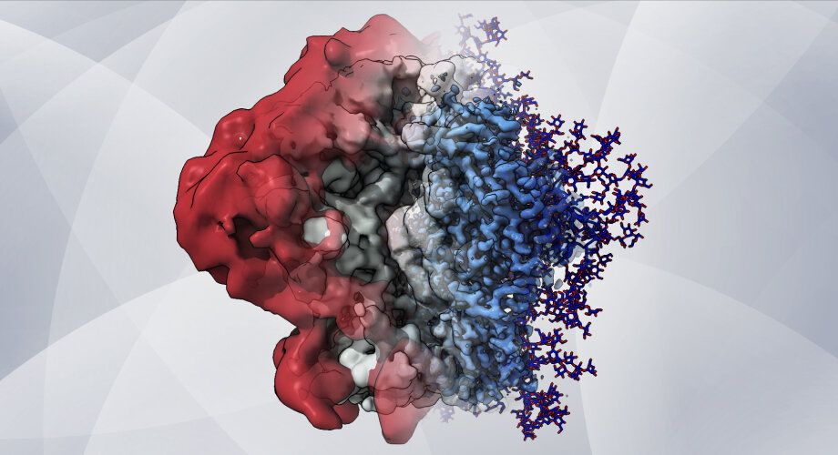
The same wave flexibility that makes these sugary molecules resistant to antibodies has made it impossible for researchers to capture them with conventional imaging at the atomic level. In the new study that appears in the Procedure of the National Academy of SciencesThe scientists developed techniques with which these elusive molecules can for the first time be mapped in detail on the surface of the HIV spike protein known as “Env”.
The Scripps Research team worked with the laboratory of Gnana Gnanakaran, Ph.D., a Los Alamos National Laboratory staff member, which is equipped with high-performance computational resources that enabled new approaches to modeling the glycans.
The researchers combined an atomic imaging method called cryo-electron microscopy (cryo-EM) with sophisticated computer modeling and a molecule identification technique called site-specific mass spectrometry. Cryo-EM relies on averaging tens of thousands or hundreds of thousands of individual snapshots to create a clear picture. Therefore, highly flexible molecules like glycans only appear as blurring if they are displayed at all.
By integrating cryo-EM with the other technologies, the researchers were able to restore this lost glycan signal and thus map vulnerable areas on the surface of Env.
“This is the first time cryo-EM has been used along with computer models to describe the structure of the virus shield in atomic detail,” said Srirupa Chakraborty, Ph.D., co-lead author and postdoctoral fellow at the Gnanakaran laboratory at Los Alamos National Laboratory.
The new combined approach revealed the structure and dynamics of the glycans in great detail and helped the team better understand how these complex dynamics affect the features observed in the cryo-EM maps. Based on this wealth of information, the team found that individual glycans do not wiggle around on the surface of the spike protein randomly, as was assumed by chance, but rather cluster in tufts and thickets.
“There are pieces of glycans that seem to move and interact with each other,” says Berndsen. “Antibodies apparently have the ability to bind between these glycan microdomains.”
Experimental HIV vaccines are based on modified, laboratory-produced Env proteins to trigger antibody responses. In principle, the effectiveness of these vaccines depends in part on the positioning and extent of the shielding glycans on these laboratory-made viral proteins. Therefore, Berndsen and colleagues used their method to map the glycans onto a modified HIV env protein, BG505 SOSIP.664, which is used in an HIV vaccine that is currently being evaluated in clinical trials.
“We found spots on the surface of this protein that are normally covered with glycans, but not – and that could explain why antibody reactions to this spot were found in vaccination tests,” says Berndsen.
This finding and others in the study indicated that Env’s glycan shield can vary depending on the type of cell used to make it. In human HIV infection, the virus uses human immune cells as factories to replicate its proteins. But viral proteins that are used to make vaccines are usually made in other types of mammalian cells.
In another surprising discovery, the team found that all of the protein began to fall apart when enzymes were used to slowly remove glycans from HIV Env. Berndsen and colleagues suggest that Env’s glycan shield, viewed purely as a defense against antibodies, may also play a role in managing the shape and stability of Env and keeping it ready for infection.
The team expects their new glycan mapping methods to be particularly useful in vaccine development and development – not just for HIV. Many of the techniques can be applied directly to other glycan-protected viruses, such as influenza and coronaviruses, and can be extended to specific cancers in which glycans play a key role, the researchers say.
Glycans in the SARS-CoV-2 spike protein play an active role in the infection
More information:
Zachary T. Berndsen et al., Visualization of the HIV-1 Env Glycan Shield Across Scales, Procedure of the National Academy of Sciences (2020). DOI: 10.1073 / pnas.2000260117
Provided by the Scripps Research Institute
Quote: New imaging method reveals the sugary shield of HIV in unprecedented detail (2020, October 23), accessed October 23, 2020 from https://phys.org/news/2020-10-imaging-method-reveals-hiv-sugary .html
This document is subject to copyright. Except for fair trade for the purpose of private study or research, no part may be reproduced without written permission. The content is provided for informational purposes only.
These were the details of the news New imaging method reveals the sugary shield of HIV in unprecedented... for this day. We hope that we have succeeded by giving you the full details and information. To follow all our news, you can subscribe to the alerts system or to one of our different systems to provide you with all that is new.
It is also worth noting that the original news has been published and is available at de24.news and the editorial team at AlKhaleej Today has confirmed it and it has been modified, and it may have been completely transferred or quoted from it and you can read and follow this news from its main source.