Das Spike-Protein
This virus has roughly 80% of the same sequences as the previous SARS-CoV. Both bind to the same host receptor, the angiotensin converting enzyme 2 (ACE2), via the S1 subunit of the viral spike protein. This is followed by the proteolytic cleavage of the spike protein at the S1 / S2 interface, with the S2 undergoing a clear conformational change and shedding the S1 subunit, creating the post-fusion form of the spike. This makes it easier for the virus to enter the cell.
The S1 subunit has the receptor binding domain (RBD), which is targeted by many natural anti-spike antibodies, as well as many vaccines in development. This is the part of the S1 subunit that binds directly to the receptor.
In the state of prefusion, the spike structure shows more significant conformational changes in the S1 region, and this is especially the case with RBD. This can have either the “up” or “down” conformation, and only the former can bind to the ACE2 receptor.
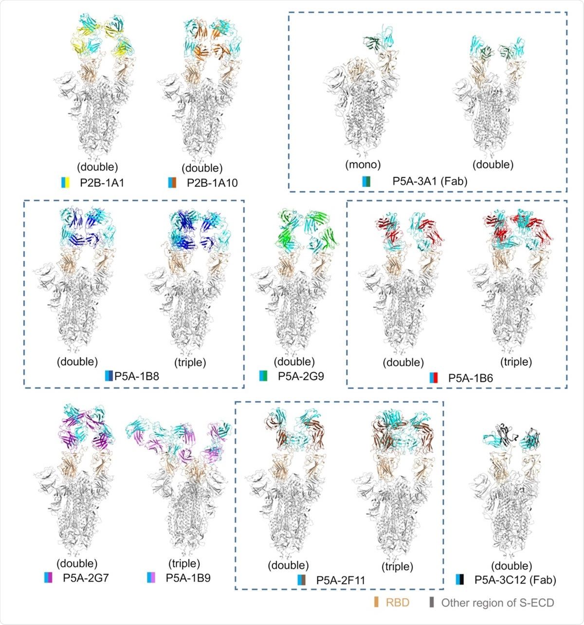
All solved structures of nAbs in complex with S-protein. The domain-colored models of all complexes are shown here. The structures that contain different numbers of the same nAb are marked with a blue dashed line. The structures are marked as mono- (1 RBD), double (2 RBDs) or triple (3 RBDs) bonds according to the number of RBDs bound with nAb.
Cryo-EM of the antibody-spike complex
The current study aims to understand how neutralizing antibodies (nAbs), which are typically divalent, bind to the trimeric spike protein. The researchers used ten cryo-EM structures of the Spike-nAb (IgG) complex, with either full-length nAb or Fab fragments or both.
Potent Human Recovery nAbs
The researchers examined ten nAbs obtained from plasma from COVID-19 patients. All were strongly neutralizing and competed with the ACE2 receptor for the binding of RBD. They also found that some nAbs slightly induced the peeling of S1 subunits, but others did not.
Structurally, they found three patterns for the nAb-spike complexes. The first included four of the ten nAbs that tied two ‘up’ RBDs. The second pattern, shown by three nAbs, had either two or three RBDs bound to the antibody in the “up” conformation. Pattern 3 was demonstrated by the most potent nAb investigated here, which bound one RBD in the ‘up’ and two in the ‘down’ conformation, with all three RBDs sharing the same binding interface.
Bivalent binding of nAbs
Previous studies have shown that divalent nAbs are more effective on viruses such as dengue and rhinoviruses compared to Fabs. The researchers therefore mapped the Fab-Spike complex separately. They found that this showed only two RBDs attached to the nAb, both in the ‘up’ conformation. With the IgG spike complex, two or three RBDs bind to the nAbs in an “up” conformation.
Despite different binding patterns, all full-length antibodies showed divalent binding of the viral spike protein.
Higher potency and S1 delivery for IgG vs. Fab shapes
They also found a difference in the orientation of the Fab-bound RBDs and the full-length IgG. Half-maximal inhibitory concentration (IC50) values were 700 times lower with the full-length antibody than with the Fab form for two of three antibodies tested, while they were over 3,000 times lower with the third.
It was found that the release of the S1 subunit at an incubation time of 120 minutes with one of the IgG nAbs was over 80% of the S protein, while the Fab form of the same antibody hardly caused any release. Other nAbs showed only a slight release.
The full-length IgG nAbs were thus able to bind more strongly and had a stronger neutralizing capacity and more significant S1 release compared to the Fab fragment binding to the spike RBD alone.
It has previously been shown that S1 shedding is a possible correlate of neutralization power, as cross-reactive nAbs against SARS-CoV and SARS-CoV-2 are typically directed against RBD, prevent ACE2 binding and promote S1 shedding. However, since the native spike protein is present as a homotrimer, it is not known whether all S1 subunits need to be rejected in order for the spike protein to change from the pre-fusion to the post-fusion form.
Comparisons of antibody binding epitopes
The structure of the Fab and scFv was designed for 8/10 and the remaining 2/10 antibodies, respectively. This indicated three groups of antibodies related to the epitopes and the angles at which they bound to the RBD.
In the first group of 7 nAbs, there was a high degree of overlap between the receptor binding motif (RBM) on the RBD and the epitopes. These antibodies recognized 8-15 of the 17 epitope residues that mediate binding to the ACE2 receptor. The angles at which the antibodies come into contact with the RBD are also comparable between nAbs in this group.
The current study confirms that of the 17 anti-SARS CoV-2 nAbs in the Protein Database (PDB) database, Group 1 nAbs are the most common.
Group 2 contains only one nAb, the most effective nAb, which was tested against both the wild type and the pseudo-SARS-CoV-2 in this study. This antibody shows pattern 3, which binds to the RBD. The direction of the RBD approach differs from that of Group 1 nAbs.
Meanwhile, the group 3 nAbs bind to the distant loop of the RBM and overlap little with the RBM alone, with a unique binding mechanism.
Of the three subgroups in Group 1, the first two induce the release of over 77% of S1, but this ability is much weaker in Subgroup 3 and Groups 2 and 3 at ~ 44% to ~ 57%. “The S1 shedding ability of the nAbs can be facilitated by the large overlap with RBM. ”
Subgroup 3 of group 1 nAbs shows a rotation of the long axis of the Fab compared to the antibodies in subgroup 1 and 2. It is possible that these antibodies can only cause a detachment of the S1 if they can reach a certain bond angle with the RBD Subunit.
Again, the presence of the Fc region in the antibody-virus complex can increase the immune response by enabling T-cell binding via their Fc receptors.
Implications
This indicated that full-length antibody binding uses a different mode and causes more RBDs to flip into the “up” conformation than the Fab form. The former includes divalent and the latter monovalent bond. The divalent bond is more effective in inhibiting virus entry and is associated with a more significant release of the S1 subunit.
Previously, the same researchers demonstrated the dimeric nature of the ACE2 receptor. In this case, each of the ACE2 molecules can bind one of the RBDs in the S-trimer, causing the bound RBD to flip up. A steric hindrance probably prevents the simultaneous binding of the two ACE2 molecules in the dimer to different RBDs of the S-trimer and thus their “unfolding”. This would require more than one ACE2 receptor to bind.
While previous studies showed many complex Spike-nAb structures, most of them used the Fab form of the antibody, especially when X-ray crystallization was used for structure visualization. The current study shows that the full-length IgG is more physiological and should be preferred for structural analysis to shed light on the actual structure and function of nAbs that target SARS-CoV-2.
* Important NOTE
bioRxiv publishes preliminary scientific reports that are not peer-reviewed and should therefore not be considered conclusive, guide clinical practice / health-related behavior, or are treated as established information.
These were the details of the news Human divalent SARS-CoV-2 neutralizing antibodies are more effective for this day. We hope that we have succeeded by giving you the full details and information. To follow all our news, you can subscribe to the alerts system or to one of our different systems to provide you with all that is new.
It is also worth noting that the original news has been published and is available at de24.news and the editorial team at AlKhaleej Today has confirmed it and it has been modified, and it may have been completely transferred or quoted from it and you can read and follow this news from its main source.